Webinar - Evaluating Spectral Cytometry for Immune Profiling in Viral Disease

Webinar Summary
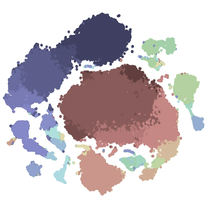
In conventional fluorescence cytometry, each fluorophore in a panel is measured in a target detector, through the use of wide band-pass optical filters. In contrast, spectral cytometry uses a large number of detectors with narrow band-pass filters to measure a fluorophore’s signal across the spectrum, creating a more detailed fluorescent signature for each fluorophore. The spectral approach shows promise in adding flexibility to panel design and improving the measurement of fluorescent signal. However, few comparisons between conventional and spectral systems have been reported to date. Here we present our findings comparing conventional and spectral approaches to cytometry—including comparisons of compensation and unmixing—and evaluate the use of spectral cytometry for immune profiling in viral diseases.
Details
Links
Time & date
- Seattle: Wed 21-Apr 1 pm
- New York: Wed 21-Apr 4 pm
- London: Wed 21-Apr 9 pm
- Zurich: Wed 21-Apr 10 pm
- Perth: Thur 22-April 4 am
- Sydney: Thur 22-April 6 am
Learning Objectives
- Gain an understanding of the essential differences between conventional and spectral approaches to cytometry.
- Appreciate the differences between compensation and spectral unmixing.
- Consider applications for spectral cytometry in the context of immunological studies.
Who Should Attend
- Researchers and technical staff who utilize flow, spectral, or mass cytometry in their work.
About the Presenters
|
Dr. Paula NiewoldPostdoctoral Researcher, Department of Infectious Diseases, Leiden University Medical Centre Dr. Paula Niewold is an immunologist currently working as a postdoctoral researcher at the Department of Infectious Diseases at the Leiden University Medical Centre. She is interested in host-pathogen interactions and how they impact the outcome of disease. She has studied these interactions in models of cerebral malaria, West Nile virus encephalitis, psoriasis, and tuberculosis using high-dimensional flow, mass, and imaging mass cytometry. She is an ISAC Marylou Ingram Scholar.
|
|
Dr. Thomas AshhurstImmunologist and High-Dimensional Cytometry Specialist, Sydney Cytometry Facility, University of Sydney Dr. Thomas Ashhurst is an immunologist and high-dimensional cytometry specialist with the Sydney Cytometry Facility at the University of Sydney. He develops and applies a range of single-cell cytometry technologies and computational analysis tools to map dynamic immune responses over time, space, and disease. In particular, he applies these approaches to the study of immunology and infectious disease, including emerging pathogens such as COVID-19, Zika virus encephalitis, and West Nile virus encephalitis. He is an ISAC Marylou Ingram Scholar.
|